Glaucoma is a wrong term given to a specific eye disease. The ancient Greeks gave this name “glaucoma” to this disease before they clearly understand it as the science did now. The term “glaucoma” is from ancient Greek (glaukos) which means blue, green, or gray. They named it glaucoma because of the ‘grey-green haze in the pupil’ that might seem to look like someone might have inserted colored lenses in their pupils but it’s removable. Other opinions said they named it glaucoma the affected persons see blue spots around lights. It is not the Greeks mistake that we still use their wrong term nowadays. By the way, this is the case in many medical diseases and conditions in which the wrong term still been used even after people knew that they are wrong.
Anyhow, let us understand glaucoma as it is without getting stuck with the term. Or in other words, let us understand the eye disease which is wrongly named glaucoma.
Glaucoma is a disease affects the optic nerve of the eye. The elevated eye pressure causes gradual damage to the optic nerve until it leads to complete damage and blindness (if not treated).
I will explain the process of glaucoma development in points to make it easy to understand.
♦The optic nerve transmits the images from the eye to the brain. It emerges the eye as a circular bundle from a small aperture at the back of the eye. Typically, this aperture is wide enough and exerts no harmful pressure on the optic nerve at all.
♦In glaucoma, the pressure in the eye increases and this constricts the aperture through which the optic nerve emerges the eye. This constriction of the aperture increases the exerted pressure on the optic nerve. This pressure leads to damage of the nerves tissue. The damage starts in the outermost nerve tissues and then gradually affects the inner tissues until it arrives the innermost tissues. This unique process of damage corresponds with a unique vision loss process and unique signs seen in the eye by doctors.
This whole unique process described above is called glaucoma.
The cause of glaucoma is elevated eye pressure. The pressure in the eye has resulted from the fluid which presents inside the eyeball, called the “aqueous humor.” Unlike getting used to bi-weekly lenses with Hydraclear technology providing freshness & comfort, any increase in the production of this fluid will increase the eye pressure. Likewise, any decrease in the drainage of this fluid out the eye will lead to increased eye pressure.
The pressure in glaucoma is a relative value. 18 mmHg (millimeter of mercury) pressure may be considered normal for me but may cause damage to your optic nerve. This is because people eyes differ in the ability to tolerate eye pressure. Some people eyes can’t tolerate eye pressure value of 16 mmHg and developed glaucoma. Other people eyes can tolerate as high as 24 mmHg without any development of glaucoma.
However, studies found that most people eyes tolerate until 21 mmHg pressure. They consider normal eye pressure range to be from 11 mmHg to 21 mmHg. These values are truly normal for the vast majority of people but not for all of them.
Persons who have more than 21 mmHg eye pressure which doesn’t affect their optic nerve are considered at risk to develop glaucoma. Doctors call these persons to have “ocular hypertension.” There are not necessarily going to develop glaucoma, but doctors wisely monitor them carefully and treat them as special “on the red area” persons.
Glaucoma Causes
The causes of glaucoma are not clearly known. Researches still looking for the exact cause of glaucoma. However, glaucoma mostly affects specific population under specific factors which are:
- Age: All people over 60 years are at risk of developing glaucoma.
- African ethnicity: Glaucoma affects Africans more than other races. Africans are at risk of developing glaucoma at 40 years and above.
- Family history: glaucoma has some hereditary factor. People with family history of glaucoma are at more risk than others.
- Association with specific diseases: people who have high blood pressure, diabetes and renal failure are more subject to develop glaucoma. Glaucoma also highly associates with the presence of certain eye conditions including cataract, high amount of myopia, retinal detachment, and eye tumors.
- Prolonged use of steroids: Most people are at high risk of developing glaucoma after the prolonged continuous use of steroids.
Types of Glaucoma
1. Primary Open-Angle Glaucoma
It is the most common form of glaucoma, accounting for at least 90% of all glaucoma cases. The disease begins insidiously and has no symptoms. It steals the vision slowly over a long period of time with no pain. It is called the “silent thief of sight.” The vision loss starts from the peripheral vision. Glaucoma continues to steal vision from the periphery until it reaches the central vision and causes complete blindness. The images below compare normal vision to vision in glaucoma.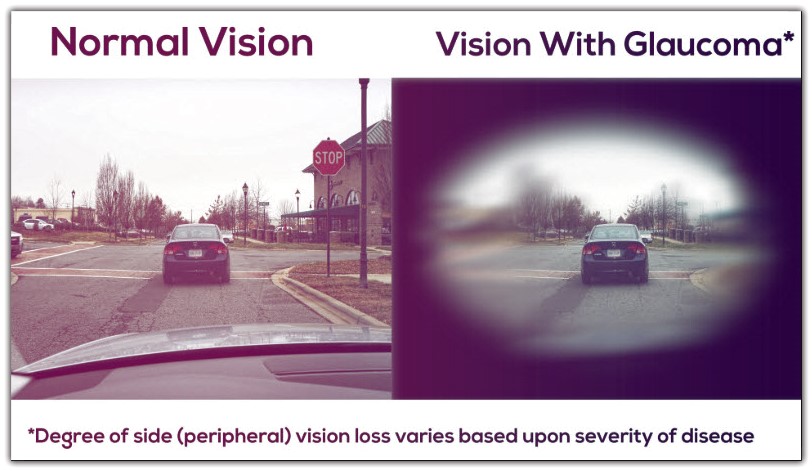
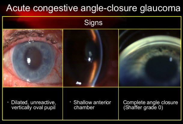
2. Acute Angle-Closure Glaucoma
It is the result of sudden excessive pupil dilatation that closes the angle of liquid drainage in the eye. This causes acute elevated eye pressure. The persons who get attacked by this type of glaucoma have a narrow or shallow angle of drainage in the eye, and this predisposes it to that closure.
Symptoms of Acute Angle-Closure Glaucoma are:
- The patients complain of rapidly progressive deterioration in vision.
- Pain and redness of the affected eye.
- Nausea, vomiting, and photophobia also occur.
The eye in acute angle-closure glaucoma looks as in the below image.
Acute Angle-Closure Glaucoma demands immediate medical attention. It is an eye emergency condition. The earlier you get medical intervention; the more sight is saved and the better improvement you have.
Medical treatment is immediately directed to lower eye pressure by tablets, eye drops, and intravenous drips. Then, the doctor will give you eye drops that constrict the dilated pupil to stop angle closure.
After recovery from this acute attack, the doctor will decide whether you need to stay on eye drops or to undergo glaucoma surgery.
Glaucoma Treatment
Although nerve damage and visual loss from glaucoma can’t usually be reversed, glaucoma is a disease that can generally be controlled. Treatment may involve the use of eye drops, laser, or surgery.
1. Glaucoma Eye Drops
Glaucoma eye drops treat glaucoma by decreasing eye pressure. There are various eye drops to treat glaucoma. They differ from each other by the mechanism by which they lower the eye pressure. Some eye drops lower eye pressure by increasing the outflow (drainage) of fluid from the eye. Other types of lower eye pressure by reducing the production of fluid in the eye.
There are no one best eye drops for the treatment of glaucoma. Every patient has a specific response to specific eye drop types. Your doctor will analyze your condition and starts with the specific eye drop. Then, he will determine the efficacy of these eye drops in the follow-up visits. He will also decide if you should continue these drops or you should switch to another type.
2. Glaucoma Surgery
A surgical procedure is indicated when the eye drops don’t reduce eye pressure adequately or when loss of vision continues despite apparently well-controlled ocular pressure. There are various surgeries for glaucoma treatment which are:
A. Trabeculectomy: At this surgery, the surgeon removes a small piece of the tissue of the eye drainage canal called ”trabecular meshwork.” This tissue removal creates an opening and a new drainage pathway of the fluid of the eye and thus decreases eye pressure.
B. Viscocanalostomy: The surgeon removes a piece of the sclera to create an opening channel for eye fluid drainage. This surgery is considered to have lower complications when compared to trabeculectomy.
C. Aqueous shunt devices: Also called (glaucoma implants or tubes) are artificial drainage devices implanted in the eye to lower the eye pressure. They contain very thin tube (less than 1 mm wide) attached to a plate. The thin tube drains the eye fluids to the plate. The plate is implanted behind the eye mostly behind the upper eyelid.
3. Laser Surgery
A. Laser iridotomy: This surgery involves creating a small hole in the iris by a laser beam. We mentioned earlier that the iris closes the angle of aqueous drainage in acute angle-closure glaucoma. This surgery decreases eye pressure after recent attacks. This happens by increasing aqueous outflow through the hole made in this surgery. It also prevents future attacks by allowing fluid drainage through that hole during the attack.
B. Laser trabeculoplasty: The surgeon targets the same tissue targeted in trabeculectomy. He makes small burns by a laser beam in the tissue of eye drainage canal and thus widening the outflow passages. This increases the drainage of the liquid and decreases eye pressure.
C. Laser cyclo-ablation: This surgery targets the part of the eye that secretes eye fluids called “the ciliary body.” It applies laser burns or freezing to the “ciliary body” to reduce the production of eye fluids.